See a 3D rotating view of this image.
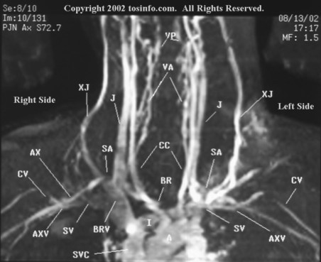
The 2D Time-Of-Flight MRA/MRV displays in high
resolution the arteries and veins which supply the brachial
plexus. The amount of blood flow is proportional to signal
intensity (which correlates to proton density). Gray, hazy
areas indicate diminished flow (compression).
In this view, the patient's arms are in the neutral position,
which is at the sides. Notice that compression of the the
nutrient blood vessels is evident even when the patient is
lying down in the neutral position. Observe the development
of collateral circulation around the compressed vessels on
both right and left sides (cloudy signal areas surrounding
the major vessels). The left axillary vein (AXV)
exhibits markedly diminished venous return as compared to
the right axillary vein (AXV).
On the left side, observe the high signal intensity of the left external jugular vein (XJ) as it enters the compressed subclavian vein (SV). The jagged irregular appearance of the left lateral internal jugular vein (J) is due to the valves within the thoracic lymph duct. Since the main thoracic lymph duct is on the left, the left side appears more jagged than the right. Observe the sharp decrease in venous return of the axillary vein (AXV) lateral to the left cephalic vein (CV) junction. The diminished venous return from within the left axillary vein (AXV) is displayed as the cephalic vein (CV) joins the subclavian vein (SV), lateral to the external jugular vein (XJ).
On the right side, observe the compression of the second division of the right subclavian artery (SA), mild effacement of the right axillary artery (AX), and costoclavicular compression of the axillary vein (AXV) lateral to the third division of the right subclavian artery. There is mild compression of the right subclavian vein (SV), medial margin to the external jugular vein (XJ). The right external jugular vein (XJ) enters the posterior margin of the brachiocephalic vein (BRV). The mild backward displaced second division of the subclavian arteries (SA both sides) reflects rounding of the shoulders.
A few labels have been placed on the image to assist with identification of landmark anatomy. The posterior vertebral veins (PV), vertebral arteries (VA), common carotid arteries (CC), brachiocephalic artery (BR), innominate vein (I), aorta (A), and superior vena cava (SVC) are labeled for reference.
A different perspective is obtained by viewing the
negative image, where the black/white color map
is reversed (i.e., black becomes white, and white
becomes black).
